Spina bifida (SPY-na BI-fi-da), a Latin term that means “split spine,” is a birth defect that occurs when a section of the baby’s spinal column does not form properly. Spina bifida treatment is available at Midwest Fetal Care Center. The Midwest Fetal Care Center is a collaboration between Allina Health and Children’s Minnesota, bringing together a multi-disciplinary team of highly trained maternal-fetal medicine experts from Allina Health and pediatric and neonatal specialists from Children’s Minnesota.
During normal fetal development, the neural tube — the hollow structure that eventually forms the baby’s brain and spinal cord — closes completely by the end of the first month of pregnancy. This closure creates a favorable environment for the formation of the complex structure and nerve connections that permit normal movement and sensation. It also protects — along with the subsequent development of a layer of skin and muscle — the spinal cord and nerves from being exposed to amniotic fluid, the liquid that surrounds the unborn baby during pregnancy (Figure 1).
Figure 1
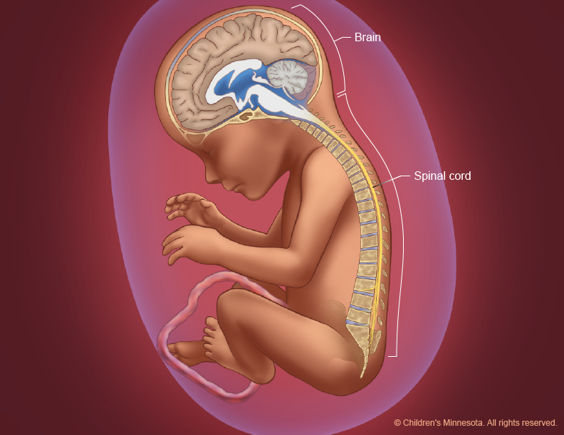
During normal fetal development, the neural tube closes completely by the end of the first month of pregnancy. The unborn baby’s muscle and skin then protect the spinal cord and nerves from being exposed to amniotic fluid.
In cases of spina bifida, however, a portion of the neural tube fails to form properly and a gap appears along the spinal column (Figure 2). In the condition’s mildest form — spina bifida occulta (oh-CULT-uh) — the gap is small and unexposed, causing fewer problems or symptoms. In other cases, however, not only does the tube fail to close properly, it also exposes the neural elements — the nerves and the spinal cord — to amniotic fluid. Generally, when doctors refer to spina bifida, they are referring to these open forms of the condition.
Figure 2

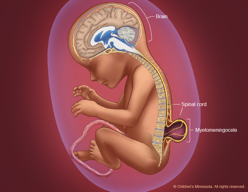
Sometimes, the protective membranes around the spinal cord push out through the gap and form a sac-like structure (Figure 3). If the sac contains the baby’s cerebrospinal fluid (the clear liquid that surrounds and cushions the brain and spinal cord), but the normal spinal cord remains in place, the condition is known as meningocele (meh-NIN-go-seal). If the spinal cord and nerves protrude into the sac, the condition is called myelomeningocele (MY-uh-lo-meh-NIN-go-seal), or spina bifida cystica (CIS-tick-uh). In some cases, the neural elements are completely uncovered, resulting in a condition known as myeloschisis (my-uh-lo-SKI-sis), the most severe form of spina bifida.
Figure 3
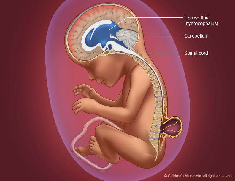
During fetal growth, the lower portion of the brain of a baby with spina bifida may shift from its normal position (Figure 4) and protrude downward into the upper spinal canal (Figure 5). This abnormal positioning — known as a Chiari II (key-AR-ee two) malformation — eventually blocks cerebrospinal fluid from leaving the brain. The result is an excess accumulation of fluid in the brain, a condition known as hydrocephalus (hy-dro-SEPH-a-lus) (Figure 6).
Figure 4
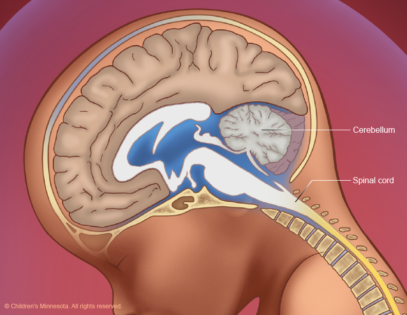
Figure 5
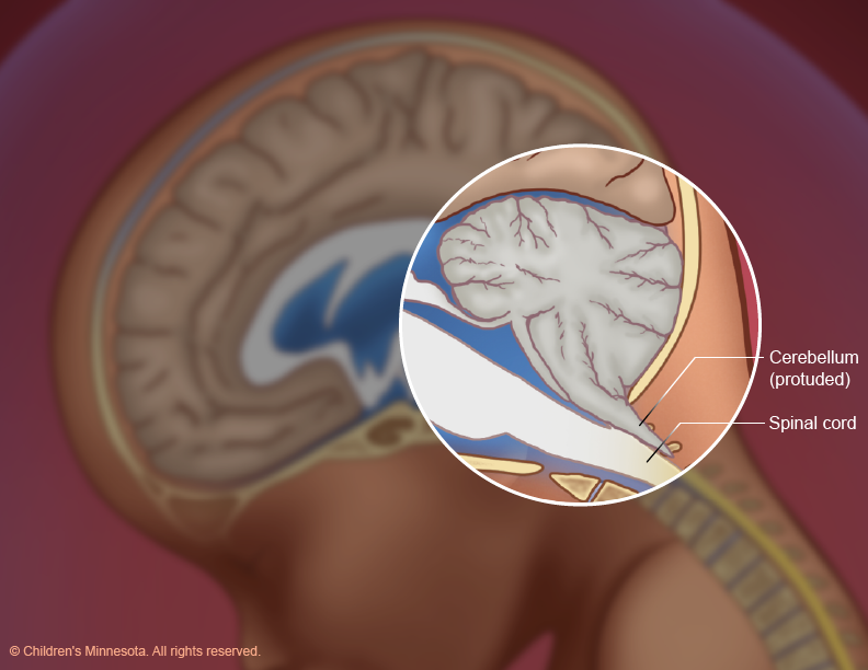
Figure 6
All these developments can result in progressive and permanent injuries. Babies born with spina bifida often have major disabilities, including weakness or paralysis of the legs and feet, bowel and bladder dysfunction, and learning problems.
Who will be on my care team?
At Midwest Fetal Care Center, a collaboration between Children’s Minnesota and Allina Health, we specialize in individual attention that starts with you having your own personal care coordinator to help you navigate your baby’s complex treatment process. We use a comprehensive team approach to spina bifida. That way, you are assured of getting the best possible information by some of the most experienced physicians in the country. For spina bifida treatment, your care team will include a maternal-fetal specialist, a pediatric surgeon, a neonatologist, a geneticist, a nurse specialist care coordinator, a fetal care clinical social worker and several other technical specialists. This entire team will follow you and your baby closely through the evaluation process, and will be responsible for designing and carrying out your complete care plan.
What causes spina bifida?
Spina bifida is the most common birth defect involving the central nervous system, occurring in about 1 in 3,000 live births. It’s believed to be caused by a combination of genetic and environmental factors. Women who have had one child with spina bifida have a 4 percent chance of having another child with the condition.
How is spina bifida diagnosed?
Spina bifida is readily detected by ultrasound. The condition is often first suspected, however, when a routine blood test — the maternal serum alpha fetoprotein (MSAFP) screening — produces abnormal results early in pregnancy. If spina bifida symptoms are suspected, your doctor will refer you to a fetal care center, such as the Midwest Fetal Care Center, for a diagnostic evaluation. At the Midwest Fetal Care Center, our evaluation process is comprehensive. It includes high-resolution fetal ultrasonography, fetal magnetic resonance imaging, amniocentesis testing and fetal echocardiography.
What is maternal serum alpha fetoprotein (MSAFP) screening?
This is a blood test that measures the level of alpha-fetoprotein (AFP) in the mother’s blood during pregnancy. AFP is a protein produced by the baby. A small amount of it normally crosses the placenta and enters the mother’s bloodstream. The most common reason for abnormal levels of AFP in the mother’s blood is a mistaken due date, but such abnormalities can also be a sign of a birth defect, including spina bifida. MSAFP screening is part of the routine screening that that women are offered starting the 15th week of their pregnancy.
What is high-resolution fetal ultrasonography?
If MSAFP screening indicates abnormal AFP levels, your doctor will suggest a high-resolution fetal ultrasonography exam. This is a non-invasive test performed by one of our ultrasound specialists. The test uses reflected sound waves to create images of the baby within the womb, including the baby’s spine. The images can help detect signs of abnormal openings in the spine.
What is fetal magnetic resonance imaging (MRI)?
Fetal MRI is another non-invasive test. It uses a large magnet, pulses of radio waves and a computer to create detailed images of your baby’s spine and other structures while in the womb. This procedure involves both mom and baby being scanned while partially inside our MRI machine. The test is a bit loud, but it takes about 30 minutes and is not uncomfortable.
What is amniocentesis?
If your baby is found to have spina bifida, your doctor will offer you an amniocentesis test to see if your baby has any associated genetic or chromosomal condition that would exclude him or her from being a candidate for spina bifida surgery in utero. This test is a necessary part of the evaluation process when prenatal surgery is being considered. For this procedure, a small sample of fluid will be removed from the amniotic sac surrounding your baby. The amniotic fluid will contain cells from your baby, and in those cells will be your baby’s chromosomes for us to analyze. The procedure is straightforward and can be done in our clinic. It requires placing a small needle through the abdomen and into the amniotic sac to obtain the fluid sample. Test results will take several days for our laboratory to process.
What is fetal echocardiography?
Fetal echocardiography (“echo” for short) is performed at our center by a pediatric cardiologist (a physician who specializes in fetal heart abnormalities). This non- invasive, high-resolution ultrasound procedure looks specifically at how the baby’s heart is structured and functioning while in the womb. This exam is important because babies with spina bifida may be at increased risk of heart abnormalities. A fetal echocardiogram is also a necessary part of the evaluation process when prenatal surgery is being considered.
How is spina bifida treated?
Spina bifida treatment depends on many factors, including the severity of the condition. At Midwest Fetal Care Center and Children’s Minnesota, we offer all spina bifida treatment options available for the care of babies with spina bifida, including prenatal and postnatal surgery. Our multidisciplinary care team will help you understand the diagnosis of spina bifida so that you can make the best treatment decision for your child.
Prenatal surgery for spina bifida
What is prenatal surgery for spina bifida?
Spina bifida requires surgery to close the spine and stop the injury to the exposed nerves. Due to remarkable medical innovations, myelomeningocele (MMC), the most common form of spina bifida, can be treated prenatally through a surgical procedure known as MMC repair.
Until very recently, the only type of prenatal MMC repair was open fetal surgery, a procedure that requires an incision through the mother’s lower abdomen and an additional incision into the uterus to expose the fetal spinal defect. Recent surgical advances now make it possible, however, to sometimes repair myelomeningocele with a less invasive technique: fetoscopic MMC repair, which eliminates the need for a large incision in the uterus. This procedure reduces the risks associated with open fetal surgery, such as rupture of the uterus, and allows the mother the ability to have a vaginal delivery.
Midwest Fetal Care Center is one of just a few medical facilities in North America that offers both open and fetoscopic MMC repair surgery. These complex procedures require a large and skilled clinical team, such as pediatric surgeons, neurosurgeons, maternal-fetal medicine specialists, and anesthesiologists, among others.
What happens during prenatal surgery for spina bifida?
Prenatal MMC repair—whether the surgery is open or fetoscopic—is done before the 26th week of pregnancy. For both procedures, mother and baby are placed under general anesthesia so that both can be operated on simultaneously.
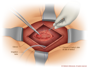
During the open procedure, a horizontal incision is made in the mother’s lower abdomen. Then, using ultrasound imaging, the surgical team makes a safe incision in the uterus, exposing the spinal defect in the unborn baby (Figure 7). With the baby’s back in view, the surgeons remove the sac, if present, and then create a watertight closure of the defect and surrounding tissue (Figure 8).
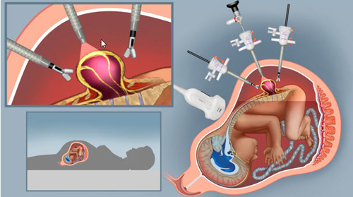
During the fetoscopic procedure, a similar incision is made in the mother’s abdomen to expose the uterus. Guided by ultrasound images, the surgical team then inserts long, thin tubes—medical devices known as trocars—into the uterus at the location of the gap in the baby’s spine. Using tiny surgical instruments, including miniature cameras, inserted through the trocars, the surgeons then repair the gap in the baby’s spine. This technique eliminates the need for a large incision in the uterus and reduces potential complications for the mother. Once the closure is completed—and watertight—the tubes are removed and the incision in the mother’s abdomen is closed.
In rare cases, the surgical team may switch from a fetoscopic to an open procedure during the surgery in order to ensure the best outcome for both mother and child.
Figure 7
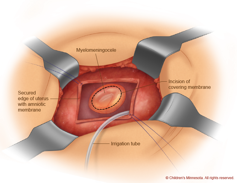
Figure 8
Fetoscopic surgery for spina bifida
During the fetoscopic procedure, a similar incision is made in the mother’s abdomen to expose the uterus. Guided by ultrasound images, the surgical team then inserts long, thin tubes—medical devices known as trocars—into the uterus at the location of the gap in the baby’s spine. Using tiny surgical instruments, including miniature cameras, inserted through the trocars, the surgeons then repair the gap in the baby’s spine. This technique eliminates the need for a large incision in the uterus and reduces potential complications for the mother. Once the closure is completed—and watertight—the tubes are removed and the incision in the mother’s abdomen is closed.
In rare cases, the surgical team may switch from a fetoscopic to an open procedure during the surgery in order to ensure the best outcome for both mother and child.
What happens to the mother and unborn baby after prenatal surgery for spina bifida?
After MMC repair surgery, expectant mothers remain in the hospital for several days. The goal of recovery is to be out of bed on the first postoperative day. Upon returning home to continue their pregnancy, expectant mothers will be advised to modify their activity. Weekly ultrasound appointments and meetings with the clinical team will also be scheduled.
After open MMC repair surgery, the goal is for expectant mothers to reach their 36th week of pregnancy, at which point the baby will be delivered via cesarean section. After fetoscopic MMC repair surgery, the goal is to have the baby born as close to the due date as possible. These babies can often times be delivered vaginally. All deliveries of babies who have had prenatal MMC repair surgery are done at The Mother Baby Center, our state-of-the-art birth center facility where you and your baby will be attended to by a highly trained, comprehensive, and multidisciplinary care team.
How long will my baby be in the hospital after birth?
Infants who have undergone prenatal fetal surgery typically remain in the hospital for a week or more after birth. The time needed in the hospital will depend on baby’s gestational age after birth and if any additional healing of the surgery site is needed. Your baby will be discharged from the hospital when he or she is observed to be feeding well, growing normally and has completed the postnatal evaluation with our team. Postnatal evaluation includes brain MRI, spine MRI, renal ultrasound, voiding cystourethrogram, video swallow study and pneumocardiogram.
What are the advantages of prenatal surgery for spina bifida?
Although prenatal MMC repair is not a cure for spina bifida, research shows that it can offer significantly better results than traditional postnatal repair. Prenatal repair can dramatically reduce the effects of spina bifida by:
- Reducing the need to insert a special tube, or shunt, into the baby’s head after birth to drain excess fluid caused by hydrocephalus. Within their first year of life, 35 percent of babies who have prenatal surgery to repair their defect will need a shunt. That compares to 80 percent of babies who have the surgery after they are born.
- Improving and reversing the Chiari II malformation (the shifting of the baby’s brain into the spinal canal). This improvement potentially reduces long-term or lifelong health problems, such as scoliosis (curvature of the spine), difficulty swallowing or breathing, and syringomyelia (fluid accumulation in the child’s spinal cord).
- Improving neurocognitive function (the child’s ability to learn).
- Significantly improving the ability of the child to walk unassisted.
Not all pregnancies are suitable for prenatal surgery. Specialists at the Midwest Fetal Care Center will meet with you to discuss the best treatment options for you and your baby.
Postnatal surgery for spina bifida
What is postnatal surgery for spina bifida?
If your baby does not have open fetal surgery, your pregnancy will go to term. Most infants with spina bifida who have not undergone surgery can be delivered vaginally. Our goal will be to have your baby’s birth occur as near to your due date as possible. Your baby will be born at The Mother Baby Center at Abbott Northwestern and Children’s Minnesota in Minneapolis or at The Mother Baby Center at United and Children’s Minnesota in St. Paul. Children’s Minnesota is one of only a few centers nationwide with a birth center located within the hospital complex. This means that your baby will be born just a few feet down the hall from our neonatal intensive care unit (NICU). Also, many of the physicians you have already met will be present during or immediately after your baby’s birth to help care for your baby right away. Your baby will be evaluated by a neurosurgeon, for example, soon after birth, and may undergo an MRI and other tests.
When will my baby have an operation?
Babies are usually operated on to repair the spina bifida defect when they are one or two days old. The procedure is similar to the prenatal one. The surgery is done under general anesthesia. Afterward, your baby will be returned to the NICU.
How long will my baby be in the hospital?
Babies who undergo post-natal surgery for spina bifida typically remain in the hospital for a week or more after birth. Your baby will be discharged from the hospital when he or she is observed to be feeding well, growing normally and has completed the postnatal evaluation with our team.
What is my baby’s prognosis?
The prognosis for babies born with spina bifida depends on the location of their defect. Thanks to medical advances, however, children born today with spina bifida can grow up to live full, happy and productive lives. As the Spina Bifida Association points out, about 90 percent of babies born with the condition now live to be adults, about 80 percent have normal intelligence and about 75 percent play sports and participate in other recreational activities.
Will my baby require long-term follow-up?
Babies born with spina bifida require life-long, multidisciplinary care. Children’s Minnesota has a variety of skilled and experienced pediatric specialists — orthopedic surgeons, neurosurgeons, urologists, physical therapists and more — who will be dedicated to the ongoing care and treatment of your child. Learn about the Children’s Minnesota Spina Bifida Clinic.
Choosing the best for your baby and family
At the Midwest Fetal Care Center, we care for the most complicated pregnancies involving the most challenging birth defects. We perform more fetal interventions than any other center in the region. Additionally, we have performed the most successful prenatal surgeries for the treatment of spina bifida in the region. In collaboration with one of the largest children’s hospital systems in the country, our extensive team of highly skilled fetal surgeons, neurosurgeons, maternal-fetal specialists, anesthesiologists and more are dedicated to the care and treatment of children and young adults.
Contact us
Need a referral or more information? You or your provider can reach the Midwest Fetal Care Center at 855-693-3825.
