Pulmonary Empyema
What is Pulmonary empyema?
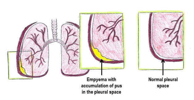
Pulmonary empyema is a buildup of pus in the pleural space (space between the lung and chest wall). It is a complication of bacterial pneumonia when the bacteria get into the pleural space and infect the tissue that lines the inside of the chest. Pulmonary empyema:
- occurs infrequently and can happen to anyone
- requires hospitalization for effective treatment
What are the signs and symptoms?
Symptoms of bacterial pneumonia include:
- Fever
- Weak cough
- No interest in activity
- Poor appetite
If empyema develops, symptoms become worse, and your child may have the following:
- Very tired
- Fever over 102°F
- Shallow and rapid breathing
Sometimes the pus pushes on the healthy lung and it becomes hard to breathe. This can cause:
- Shortness of breath with gasping
- Pale or grey color
- Difficulty walking
- Chest pain with anxiety
How is pulmonary empyema diagnosed?
Tests to diagnose pulmonary empyema include:
- Chest x-ray to confirm that an empyema is present and estimate the amount of pus in the chest
- Ultrasound of the chest to determine the thickness and amount of pus
- A computed tomography study (CT scan) may be done if there is any question about the diagnosis, but is usually not necessary
- Blood tests to check for changes due to the empyema
- A culture of the pus from the chest is the best way to identify the bacteria causing empyema
How is it treated?
- Pain medicine is used to keep your child comfortable
- Antibiotics are given for 14 days or more depending on the bacteria causing the infection
- A chest tube (a small tube inserted between the ribs through the chest wall) is placed to drain the pus from the inside of the chest (see education sheet “Chest tube”)
- The chest tube stays in place for 3 to 5 days
- Oftentimes, medicine is given through the chest tube to help dissolve and drain the thick pus
In 10-20% of patients, surgery may be needed to help drain the pus. This surgery is called a VATs procedure (Video-Assisted Thoracoscopic surgery). Several small incisions are made in the chest wall: one for a small video camera and others for instruments. Pus and blood are removed from the chest. After the procedure the child returns to the hospital room with a chest tube in place to collect any remaining pus. This chest tube stays in place for 3 to 5 days. Pain medications are given to keep your child comfortable.
What else do I need to know?
- Your child will be eligible for discharge after the chest tube is removed and he or she is able to drink fluids and take oral medications.
- Children can easily lose 1 – 5 pounds during a complicated infection and nutrition is important to help with wound healing.
- Remember, the lung is temporarily injured during pneumonia, but will heal with antibiotics and nutrition.
- Pulmonary empyema is an uncommon complication of pneumonia and rarely recurs.
- Children who experience pulmonary empyema are not vulnerable to subsequent lung infections.
- Lung function studies performed months or years after pulmonary empyema reveal that children have remarkable healing ability.
- Pulmonary empyema generally does not limit a child’s ability to participate in normal activity within one month of the event.
- Additional antibiotics for 1 or more weeks may be required.
Questions?
This information is not specific to your child, but provides general information. If you have any questions, call your clinic.
Reviewed by Kuracheck 2/2017
Back To Top
