This blog was medically reviewed by Lisa Howley, MD, FASE, FACC, director of the fetal cardiology program at Children’s Minnesota.
If you are pregnant and are told that you need a fetal echocardiogram done, it can feel a little scary, but we’re here to help. There can be a variety of reasons to have a fetal echocardiogram done, and we are here to talk you through the process, what to expect and more.
Early detection of congenital heart problems saves lives. While most heart defects can be detected while babies are still in the womb, many health systems do not have the expertise to diagnose — and treat — those conditions. But, Children’s Minnesota is different. In this blog, you’ll learn about fetal echocardiograms, the reasons you may need one, how they are done and more.
What is a fetal echocardiogram?
A fetal echocardiogram (“fetal echo” for short) is a specialized type of ultrasound that allows diagnosis of heart problems in utero. This fetal cardiac echo is a non-invasive ultrasound that is focused on evaluating the unborn baby’s heart structure and how it’s functioning while in the womb.
Detailed information from the fetal echo test enables our specialized fetal cardiology team to diagnose structural, functional and heart rhythm abnormalities prior to delivery.
If there is concern about your baby’s heart on routine prenatal ultrasound, a fetal echocardiography and consultation with a fetal cardiologist may be recommended. Fetal echos can be performed at specialized maternal-fetal medicine locations and comprehensive fetal care centers like the Midwest Fetal Care Center, a collaboration between Allina Health and Children’s Minnesota.
Reasons to have a fetal echo
There can be a wide variety of reasons your health care provider may want you to have a fetal echo ultrasound done. Here are some of the common indications for a fetal echo:
- An abnormality is identified on your routine prenatal ultrasound that raises concerns that your baby may have a heart problem.
- The pregnant mother, father of the baby, or sibling of the baby has a history of congenital heart disease, called a congenital heart defect.
- Your baby has a suspected chromosome or genetic abnormality.
- You have a twin pregnancy where the two unborn babies share a placenta.
- The pregnant mother has pre-existing Type 1 or uncontrolled Type 2 diabetes.
- Your baby has a separate non-heart-related problem, such as a defect in the abdominal wall, a lung problem, a defect in the diaphragm, a brain malformation or a kidney abnormality.
At what point in pregnancy is a fetal echo done?
You may be wondering, at what point will an echo in pregnancy be done? A fetal heart ultrasound can be done any time after the 13th week of pregnancy. As mentioned above, early detection of congenital heart problems saves lives so, if there’s a concern that your baby may have a heart problem, we encourage a prenatal echo as early as possible.
As one of the only comprehensive programs in the region equipped for both prenatal and postnatal cardiovascular care, we continually aim to improve outcomes for our tiniest heart patients. For families diagnosed with a fetal heart abnormality, our fetal cardiology team works alongside your health care provider to develop an individualized care and treatment plan.
Learn more about the largest regional fetal cardiology program with 2,000+ fetal echocardiograms performed annually.
How is a fetal echocardiogram done?
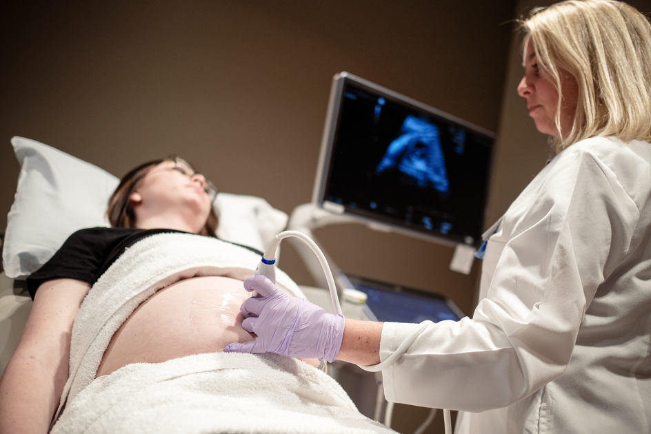
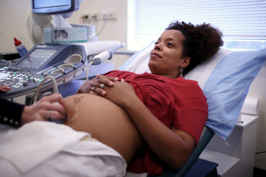
A fetal echocardiogram for pregnancy will typically be similar to your previous ultrasounds for pregnancy. A specialized ultrasound technician, called a sonographer, performs the fetal echo. The sonographer will place ultrasound gel to your pregnant abdomen and then they will use an ultrasound probe to take pictures of your baby’s heart.
Following completion of the fetal echo, a fetal cardiologist reviews the images and sometimes performs a brief scan as well.
How long does a fetal echo take?
A fetal echo typically takes between 30-60 minutes but can sometimes be longer. The required time can vary depending on the baby’s position, the number of babies in the pregnancy (twins, triplets, etc.), and the images your care team needs.
What should I do to prepare?
For a fetal echo, there is no need for special preparation for this test. You don’t need to have an empty stomach or have a full bladder.
What will the results show?
Your cardiac fetal ultrasound results are provided during the same visit after the test is performed and reviewed by the fetal cardiologist. The fetal cardiologist will review the results of your fetal echo with you, explaining the normal or abnormal findings in detail and they will help arrange follow up when needed.
What are my next steps?
At the Midwest Fetal Care Center, we will work with you and your referring provider to develop a comprehensive care and delivery plan. We will fully prepare you for what to expect for you and your baby.
Fortunately, most unborn babies can tolerate a heart abnormality without needing any in-utero treatment or intervention. Plus, the majority of heart conditions are best treated after the baby is born.
But in some circumstances, fetal medical treatment or fetal surgery may be considered for life-threatening cardiac conditions. It is our goal to provide you and your family with the information you need to make an informed decision about the best treatment options.
How the experts at Children’s Minnesota and the Midwest Fetal Care Center can help
At Midwest Fetal Care Center, the fetal cardiology team collaborates with top-notch experts in maternal and fetal medicine as well as specialists in newborn and pediatric care, including neonatologists. Our team works together to coordinate all aspects of your care from diagnosis to birth planning and long-term follow-up.
Children’s Minnesota and the Midwest Fetal Care Center are here for you and your family every step of the way. We hope this article has helped you further understand the basics of a fetal echocardiogram, what to expect and how you can prepare.
Learn more about the leading provider of pediatric cardiology services.