Key points:
- Specialists from Sanford Health and Children’s Minnesota partnered to treat Amelia, an adolescent girl who was diagnosed with a rare brain tumor called pleomorphic xanthoastrocytoma (PXA).
- A vital artery was tightly adhered to the tumor, making total surgical resection more difficult. After a partial resection at Sanford, a second procedure performed at Children’s Minnesota utilized a pediatric hybrid intraoperative MRI suite, which allowed for real-time imaging and ensured complete tumor resection before leaving the operating room.
- Following the second surgery, Amelia has remained seizure-free, and her recent follow-up MRI showed no signs of residual or recurrent tumor.
Life took an unexpected turn for 12-year-old Amelia when she suffered a seizure in front of her family in February 2024. It was the most alarming sign of what turned out to be a primary central nervous system (CNS) tumor called pleomorphic xanthoastrocytoma (PXA), a very rare type of astrocytoma.
Initial brain surgery
After the seizure, Amelia’s mom Danielle took her to the Sanford Luverne Medical Center in their hometown of Luverne, Minn., where a CT scan revealed a tumor in her brain. She was then transferred to Sanford Children’s Hospital in Sioux Falls, S.D.
A few days later, Amelia had a right frontal craniotomy. Shawn Vuong, MD, neurosurgeon at Sanford, removed as much of the tumor as possible before he had to stop because a branch off the middle cerebral artery (MCA) was tightly adhered to the tumor. The artery is particularly important as it supplies the part of the brain responsible for movement on the opposite side of the body. Injury to the artery during surgery, such as bleeding or a lack of blood flow, would have significantly impacted Amelia’s daily life.
There are two grades of PXA tumors – Grade 2 are low-grade tumors and Grade 3 are malignant, fast-growing tumors. A biopsy of the tumor was taken during surgery which confirmed Amelia had a Grade 2 PXA located in the front area of the brain.
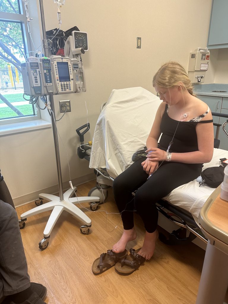
Since some of the tumor remained following surgery, Amelia went through several rounds of chemotherapy. Unfortunately, the tumor was not responding to the treatment. In addition, she was still having seizures that felt like a tingling sensation in her hands and legs two to three times a day, despite being on medication.
Amelia’s case referred for second opinion
A new option for Amelia and her family came in June 2024. Catherine Nelson, DO, a pediatric oncologist at Sanford Children’s Hospital, contacted Anne Bendel, MD, research director of neuro-oncology at Children’s Minnesota, to review Amelia’s case and provide a second opinion.
Amelia’s case was reviewed at the Children’s Minnesota tumor board conference where a panel of neurosurgeons, neuro-oncologists, neuro-radiologists, neuro-pathologists and geneticists review a patient’s clinical, imaging, surgical and pathology histories. In addition, Meysam Kebriaei, MD, medical director of neurosurgery, reviewed Amelia’s case directly with Dr. Vuong to learn what he encountered during surgery.
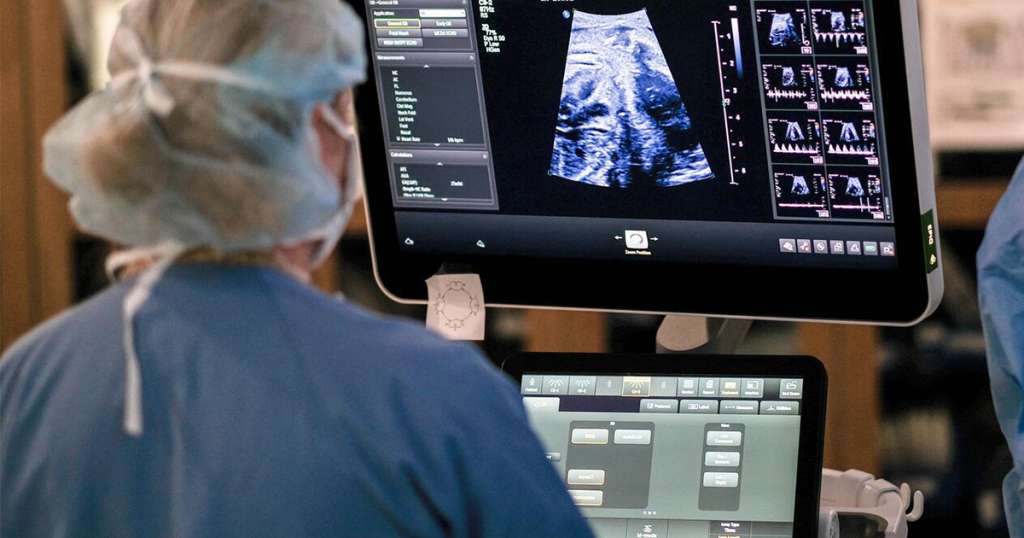
After the detailed review, Dr. Kebriaei and the tumor board recommended that the neuro-oncology and neurosurgery teams at Children’s Minnesota could provide Amelia with the opportunity for the best outcome – total resection of the tumor. Among the deciding factors was the pediatric hybrid intraoperative MRI suite – the first pediatric neurosurgery suite of its kind in North America. It’s equipped with both moving-scanner and moving-patient MRI technology. The iMRI takes crystal-clear images mid-procedure, allowing care teams to make enhanced clinical decisions to improve patient outcomes.
Coordinated care with referring providers
Dr. Nelson shared the news with Amelia’s family and soon after they had a virtual consultation with Dr. Kebriaei. During the consultation, Dr. Kebriaei explained how the iMRI suite would allow them to confirm if any of Amelia’s tumor remained before leaving the operating room. Once all their questions were answered, the family felt confident moving forward with a second surgery.
After the referral, Dr. Bendel and the Children’s Minnesota neuro-oncology team and Dr. Nelson and the oncology team at Sanford Children’s Hospital remained in close contact throughout to help coordinate Amelia’s care pre- and post-surgery as well as planning her ongoing follow-up and management in the future.
“We’re fortunate to have the collaboration with Dr. Bendel and the team at Children’s Minnesota on cases like Amelia’s that require a highly specialized level of care,” said Dr. Nelson. “After the referral, we truly share in the care of these patients, with each side providing updates to the other.”
Second brain surgery
In September, Amelia and her parents came to the Children’s Minnesota hospital in Minneapolis for her second right frontal craniotomy. The delicate procedure led by Dr. Kebriaei took approximately nine hours. An iMRI taken in the operating suite post-tumor resection showed no evidence of residual tumor.
“Surgical removal of tumors like Amelia’s are known to be more difficult, which is why the iMRI suite and real-time imaging is so vital in cases like hers,” said Dr. Kebriaei.
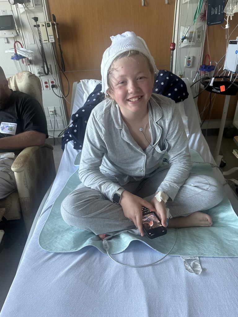
Amelia returned home to her family a few days later and has remained seizure-free since then. She continues to receive collaborative follow-up care at Sanford and Children’s Minnesota. Her three-month follow-up MRI in December showed no signs of residual or recurrent tumor. In that same month, she was also cleared to resume figure skating, a passion she has pursued for four years.