How is renal agenesis diagnosed?
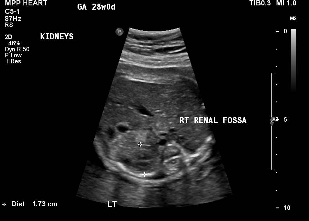
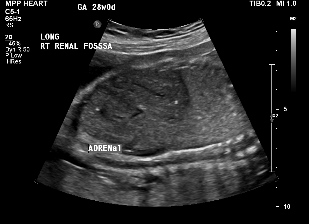
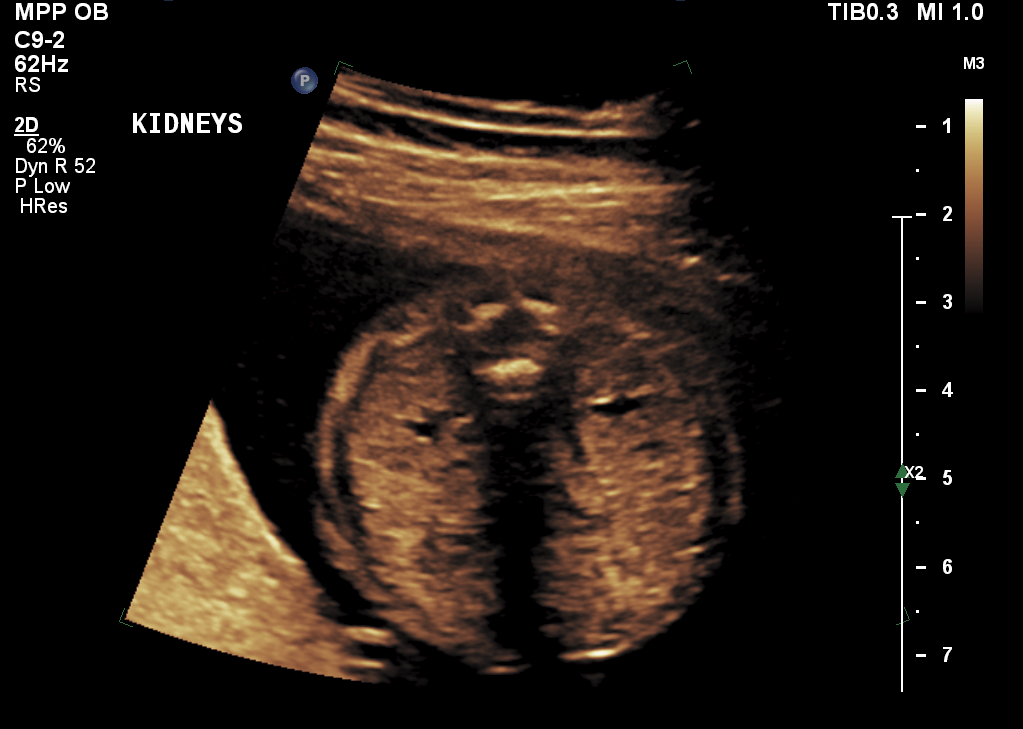
Renal agenesis is usually identified during a routine prenatal ultrasound. The initial telltale sign of the condition is an empty space (renal fossa) where the kidney should be (Figure 2). Also, the adrenal gland — a pyramid-shaped structure usually attached to the top of a kidney — will appear flat on the ultrasound, as if it were “lying down” (Figure 3).
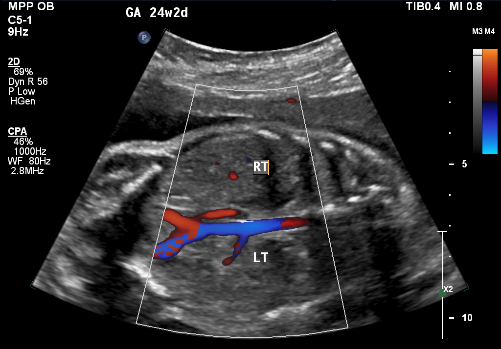
Kidneys can sometimes develop in the wrong place within a baby’s body, a condition known as an ectopic kidney. To rule out that possibility, your doctor will perform an ultrasound scan, which uses sound waves to identify your baby’s anatomic structures. It can also measure the flow of blood through the baby’s blood vessels. Your doctor will look at the ultrasound images to attempt to identify renal (kidney) tissue and renal arteries, which carry blood from the heart to each of the kidneys. The absence of renal tissue, along with the absence of the renal artery on the same side (Figure 4), confirms the diagnosis of renal agenesis.
If no renal tissue is identified and the ultrasound also shows no amniotic fluid in the womb, the diagnosis is likely to be bilateral renal agenesis. The presence of renal tissue with a lack of fluid could be a sign, however, that the membrane around the amniotic fluid — the amniotic sac — has ruptured. Your doctor will either confirm or rule out this possibility by performing a pelvic examination to look for signs of pooled amniotic fluid in the vagina. Other tests may also be done to evaluate for the presence of amniotic fluid, which is usually not found in the vagina.


How is unilateral renal agenesis managed before birth?
If your baby is missing a single kidney, the prenatal care you receive will center on acquiring as much information about the baby’s condition as possible so that we can be prepared during and after delivery to provide optimal care. That includes collecting information on other possible birth defects.
We use several different techniques to gather that information, including high-resolution ultrasonography and fetal echocardiogram. If these tests reveal no birth defects other than the single missing kidney, your baby will not require any special prenatal treatment. An extra ultrasound may be done in the late second or early third trimester of your pregnancy, but otherwise you will receive routine prenatal care.
What is high-resolution fetal ultrasonography?
High-resolution fetal ultrasonography is a non-invasive test performed by one of our ultrasound specialists. The test uses reflected sound waves to create images of your baby within the womb.
What is fetal echocardiogram?
Fetal echocardiography (“echo” for short) is performed at our center by a pediatric cardiologist (a physician who specializes in fetal heart abnormalities). This non- invasive, high-resolution ultrasound procedure looks specifically at how the baby’s heart is structured and functioning while in the womb. This test is important because babies with birth defects are at increased risk of heart abnormalities.
How is bilateral renal agenesis managed before and after birth?
There are no established treatment guidelines, either before or after birth, for babies who are missing both kidneys. Parents of babies with bilateral renal agenesis have the option of not continuing with the pregnancy or, if they wish to continue with the pregnancy, of having their baby receive palliative care after birth.
Some babies with the condition may qualify for an experimental treatment, known as Renal Anhydramnios Fetal Therapy (RAFT). A clinical trial is currently underway to evaluate whether this intervention can improve survival. The trial is currently recruiting patients. Its goal is to preserve the function of the baby’s lungs before birth by making a series of injections of fluid into the amniotic sac to maintain normal fluid volumes. After birth, the baby will likely need to undergo kidney dialysis. A kidney transplant will likely also be needed at some point during the first few years of life.
During the dialysis procedure, a machine will remove waste products and excess fluid from your baby’s blood—the job that kidneys normally do. During the kidney transplant surgery, a surgeon will place a kidney from a donor into your baby’s abdomen. The kidney will then be attached to nearby blood vessels to enable it to start working.
To qualify for a RAFT clinical trial, a baby must have no other serious birth defects, normal genetic testing, and a good chance of surviving the treatment. Your baby’s doctor can counsel you on whether your baby is a candidate for this experimental therapy.


